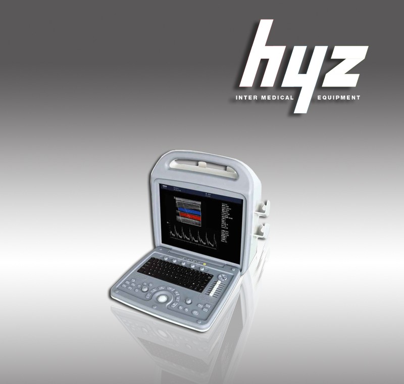
HYZ-CDU580 Portable Color Doppler Ultrasound diagnosis system
|
1.1Device Name: Full Digital Color Doppler Ultrasound |
|
1.2Device Application: abdomen,maternity, urology, superficial organization, peripheral vascular, small organ, cavity, and other interventional screening and therapeutic use. |
| 2.Technical specifications |
|
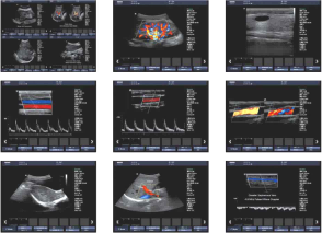
2.1full-digital color ultrasound diagnostic system, the system has the ability to upgrade 2.2Frequency Range :2.0-14MHz 2.3Probe types: convex array, linear array, heart and cavity probes 2.4Display: B / M, B / D, B / CD, B / D / CD may be the same screen display, M type and scope of PWD icon size adjustable 2.5 Frequency Probe Specifications: 2.5.1Abdomen convex array probe: 2.0 ~ 5.0MHz electronic convex array broadbandprobe frequency, the frequency range 2 - 5MHz, 3 selectable frequencies Bandwidth, scan angle: 60 degrees. 2.5.2E-broadband frequency linear array probe, the frequency range 5 - 15MHz. 2.5.3Intracavity frequency convex array broadband probe, the frequency range 4 - 8MHz, scanning angle ≥ 135 ° 2.5.4Optional pediatric cardiac probe, transesophageal probe, laparoscopic probe, tiny protruding array probe, intraoperative probes. 2.6probe array element: ≥ 128 2.7Image Mode: two-dimensional B-, M-type, pulsed PW , color CFM / power Doppler and direction of the Energy PDM 2.8 B / D use either: B / PWD, B / CD / PWD three simultaneous display 2.9Organization of second harmonic imaging, harmonic function ≥ 2 groups 2.10Resolution: Lateral ≤ 2mm, longitudinal ≤ 1mm 2.11detecting depth: ≥ 240mm 2.12system dynamic range ≥ 140dB 2.13Image playback video: frame by frame, continuous playback of ≥ 300 frame 2.14 Focus: Emission ≥ 8 segment focus, to receive: continuous dynamic variable aperture, dynamic apodization digital focusing 2.15Scan Line: Every frame linear density ≥ 400 Ultrasonic Line 2.16Measurement and Analysis 2.16.1General measurement: distance, perimeter, area, slope, heart rate, pressure, residual urine, RI, PI, flow rate, etc. 2.16.2 Special obstetrics and gynecology measuring software: fetal gestational age at due date, and estimation, fetal weight, measurement take many times average, S/D ratio, etc 2.17Character Tags: shows the date, time, patient's name, user name, etc., custom note table, probe, frequency and body marked, with a puncture and guide lines 2.18keyboard operation: Sino-British operation interface 2.19position markers: ≥ 30 Zhong with the location of the probe position markers 2.20Color Doppler display modes: speed dispersion shows that the energy shows that dispersion Show 2.21display position adjustment: linear array scanning range of interest: -20 ° - +20 ° 2.22Doppler flow velocity: The maximum blood flow velocity measurements: PWD ≥ 6m / s; flow velocity measurements: PWD ≥ 10mm / s2.26width and location of sample volume adjustment :0.5 - 20mm adjustment classification 2.27with a Doppler angle correction function for sampling and then 2.28Display: 15-inch high-resolution progressive-scan LCD displays. 2.29Probe Interface:2 units 2.30picture archiving and management 2.30.1Dynamic and static image real-time hard drive storage capabilities, the host built-in hard disk ≥ 160G. 2.30.2There was a picture online clipboard functions: real-time scan, only one button operation, can be dynamic and static ultrasound images are stored in the screen side of the clipboard, you can always transfer out of contrast observation. 2.30.3The original data acquisition and processing capabilities, can playback dynamic and static image post-processing capabilities, and can convert directly to the image avi, tif, Bmp, and other common format computer 2.31Input / Output Signal Interface: PAL-D, USB, port, etc. 2.32Supply Voltage: AC |
|
Volume (mm):580×530×400 G.W. (kg):16 N.W. (kg) |
|
Excellent image quality from advanced digital imaging technology
Ultra-wideband digital beamformer To ensure without loss of diagnostic information that effective control ultrasonic beam to eliminatethe side lobe noise,greatly enhancing the image spatial resolution and contrast ratio. Echo optimization technology to enhance the organization- high color sensitivity according to different degree of depth and fat to optimize images automatically delete ultrasound artifacts(Ghosting) Automatic color flow filter extravascular spillover,make the image more realistic,clear-cut Increased color flow (CFM) and the energy GPD sensitivity |
|
Full motion colour Doppler(CFM),colour Doppler energy diagram(CPD) Pulse Doppler(PW),
Perfect measurement package(abdominal,vascular,maternity,paediatrics and urology,etc) All clinics,adult and pediatric cardiac,abdominal,obstertrics and gynecology,urology,pediatrics,endocrine branch Peripheral vascular,small orgn(breast,eye,esophagus,muscles,etc)through the skull,urgery,puncture,etc Touch screen operation of navigation systems,simplifying the complex procedures With a fully scalable digital image management system can be supply 12V DC power |