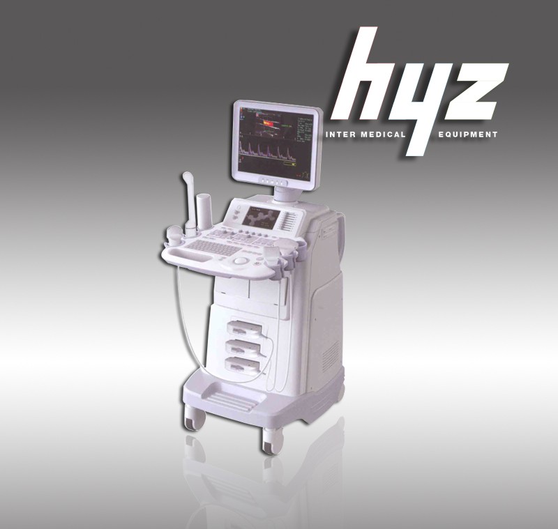
HYZ-CDU480 Full digital CFM Ultrasound Imaging System
image quality.
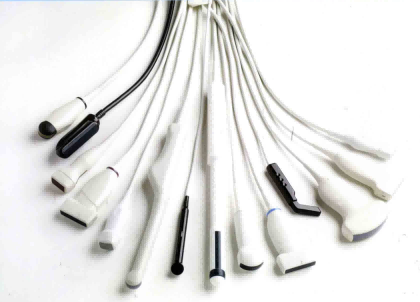
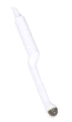
Probes and accessories for a wide group of
 |
 |
|
Support for high-resolution 4D cavity probe,scan for,and the computer storage and recovery. |
|
 |
 |
|
Abdominal probe 4D volume quickly get information. |
|
Ultrasound diagnosis system parameters |
|
1. The host system |
|
1.1 Full-digital color Doppler ultrasonic diagnostic system
1.2 Ultrasound host operating system: Windows XP operating system
1.3 Monitor: ≥ 17-inch high-definition professional medical LCD displays, with a 7-inch
widescreen touch-navigation system
1.4 Probe connector (all effectively activate) ≥ 3
1.5 2D gray-scale imaging unit
1.6 The PW pulse wave Doppler
1.7 Doppler energy and directional energy diagram
1.8 Color Doppler ultrasound diagnostic unit
1.9 The THI tissue harmonic Imaging
Full (Wide) scene imaging
Extend the (extended) pulse imaging (EPI)
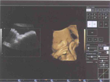
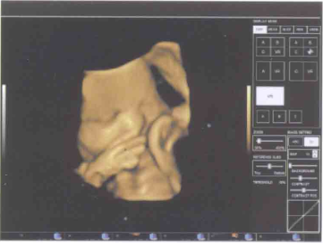
Real-time 3D/4D synchronization imaging
Trapezoidal extended imaging
Anatomical M-mode imaging
Elasticity imaging (ELASTOGRAPH)
1.10 Inverting pulse harmonic imaging
1.11 A key optimization (optimize 2D gray-scale images and PW images)
1.12 Multi lingual user interface optional, Chinese/English operation interface, support for
Chinese/English input.
1.13 Real-time synchronization unit: 2D, color Doppler, spectral Doppler / continuous
CWD at the same time the same screen real-time display.
1.14 Professional package: abdomen, obstetrics and gynecology, small parts, urology,
orthopedic surgery, cardiac1.15 Measurement: distance, area, angle, time, slope, heart rate, speed, acceleration,
spectrum tracing, resistance index and pulsate index
1.16 Gain: B / PW / CD can be separately adjusted
1.17Cine Loop ≥ 1000 frame
|
|
2. Probe configuration and specifications: |
|
2.1 Configuration: convex array probe, the high-frequency linear array probe, transvaginal
probe
2.2 convex array probe :2.5-5 .0 MHz center frequency: ≥ 3, visual adjustable; with
harmonic imaging capabilities, the harmonic frequency of ≥ 2, visual adjustable
2.3 linear probe :6.0-10 .0 MHz center frequency: ≥ 3, visual adjustable; with harmonic
imaging capabilities, the harmonic frequency≥ 2, visual adjustable
2.4 Transvaginal probe : 5.0-8 .0 MHz (scan angle ≥ 130°)
2.5 Variable frequency phased array cardiac probe: the frequency range 2 - 4MHz, 3
frequency bandwidth is optional; probe scanning angle:10°to 85°.
|
|
3. 2D imaging modes: |
|
3.1 4B imaging mode
3.2 B-type images in real time and freeze the zoom magnification of ≥ 8, 10 or more
adjustable
3.3 Grayscale: 256
3.4 Position chart: ≥ 60
3.5 Dynamic range: 0-150db, visual adjustable 10 level and above
3.6 The probing depth: ≥ 360mm
3.7 Resolution: lateral ≤ 2mm; vertical ≤ 1mm
3.8 Focus: B mode, the focus position and focus distance is adjustable
3.9 Ultrasonic power output: ≥ 10 adjustable
3.10 Preconditions: ≥ 10 species, the user can customize the conditions
3.11 Pseudo-color display: ≥ 7 species
3.12 Convex array probe: 18cm depth, full aspects, 2D frame rate≥ 30 frames / sec.
3.13 Linear array probe beam deflection
3.14 Digital beam formation: continuous dynamic focusing, variable aperture and dynamicchange trajectory
3.15 B, 2B, 4B display modes
3.16 TGC segment gain adjustment ≥ 8 segments
|
|
4. M model |
|
4.1 B / M to a variety of layout adjustable 4.2 M scanning speed is adjustable |
|
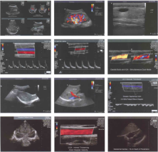
5. Color Doppler: |
|
5.1 Color gain and PRF multi-level adjustable 5.2 Visual Color frequency adjustable: ≥ 2
5.3 Sampling frame: the size and position adjustable 5.4 Color Atlas: ≥ 7
5.5 Color afterglow: ≥ 4 5.8 Color baseline adjustable
|
|
6.Color Doppler energy diagram: |
|
6.1 The direction of the energy diagram 6.2 Energy in gain: Adjustable 6.3 The energy diagram |
|
7. Doppler spectrum: |
|
7.1 Doppler spectrum of the emission frequency can be multi-file variable frequency
7.2 The measurement speed: the minimum detectable speed of 1mm / s maximum speed
measurement ≥ 25m / s,
7.3 Sample volume size: 1mm-15mm, adjustable
7.4 Sampling point of correction: -70 °to 70°
7.5 Spectrum Gain: adjustable;
7.6 Spectrum Automatic tracing and automatic measurement functions, including
automatic envelope, manual envelope, fast measurement
7.7 Tracings range to support the baseline above and below the baseline, and all tracings
in three ways
7.8 Baseline: Zero moving grading adjustable ≥ 8 levels
7.9 Scan speed: ≥ 3 adjustable7.10 Wall Filtering: ≥ 5-level adjustable
7.11 Spectrum volume size is adjustable
7.12 Spectrum envelope automatically in real time measurement and calculation
|
|
8. Iimage-text management system: |
|
8. Iimage-text management system:
8.1 Built-in storage unit: hard drive storage capacity ≥ 320G;
8.2 Built-in ultrasound image archiving and management functionality: You can edit the
diagnostic report , the ultrasound diagnostic images can be embedded in the report and
print directly
|
Excellent Image quality
Full motion color Doppler (CFM),color Doppler energy diagram (CPD) Pulse Doppler (PW),continuous Doppler (CW)
Improve the measurement package (abdominal,vascular,maternity,paediatrics and urology,etc.)
All clinics,adult and pediatric cardiac,abdominal,obstetrics and gynecology,urology,pediatrics,endocrine Branch
Peripheral vascular,small organ (breast,eye,esophagus,muscles,etc.) through the skull,surgery,puncture,etc.
Large-screen LCD display,multi-angle full range of movement,the real clinical ultrasound for the diagnosis of the first line of
Touch-screen operation of navigation systems,simplifying the complex procedures
With a fully scalable digital image management system
Ultra-wideband digital beamformer
To Ensure without loss of diagnostic information that effective control ultrasonic beam to eliminate the side lobe noise,greatly
enhancing the image spatial resolution and contrast ratio.
Improve sensitivity of colour flow CFM and energy app
Tissue echo enhancement colour optimizatim technology-high sensitivity.
According different depth and fat,thin to optimize images
Delete ultrasound ghosting automatically
Filtered blood spills outside colour flow automaticallymake the image more realistic and clear
Channel domain processing techniques
New,very different ultrasound technology platform
Full software control of the beamformer
Faster processing speed,more abundand amount of information