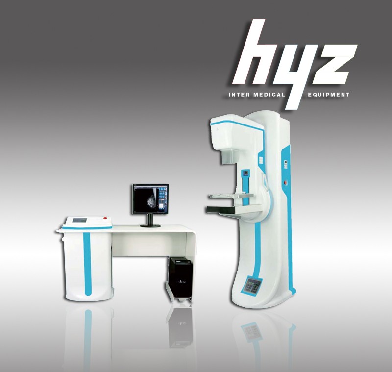
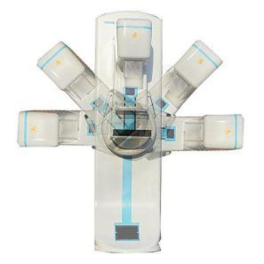
HYZ-Mega 600 Digital Mammography System
|
Item |
Length * Width *height/mm |
Weight/kg |
|
Host machine (without packing) |
2000 *560 *940 |
150 |
|
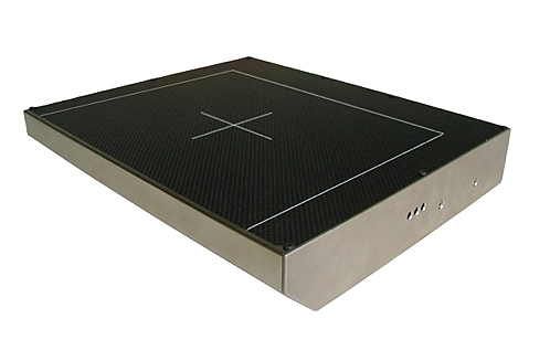
Operation platform (without packing) |
1500 *930 *500 |
85 |
|
Host machine (packing) |
2160 *690 *1150 |
210 |
|
Operation platform (packing) |
1630 *970 *830 |
140 |
I. Application
A mammogram is a special, low-dose X-ray technique used to take a picture of the breast, detecting and diagnosing any abnormal lumps or masses in breast tissue. It is one of the best tools for the early identification of breast cancer. With early identification, breast cancer can be cured while in the first stage, and recovery is more likely.
II. Specification:
III.Configuration:
|
No. |
Item |
Quantity |
|
1 |
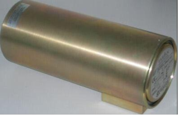
X-ray Tube |
1 |
|
2 |
X-ray Generator |
1 |
|
3 |
Gantry assembly |
1 |
|
4 |
C-ARM |
1 |
|
5 |
Bucky movement device |
1 |
|
6 |
Flat panel detector |
1 |
|
7 |
Image acquisition workstation |
1 |
|
8 |
Review work station |
1 |
|
9 |
Paddle switch |
2 |
|
10 |
Exposal switch and connected line |
1 |
|
11 |
Power wire |
1 |
|
12 |
Grounded wire |
1 |
|
13 |
Fuse |
2 |
|
14 |
Operation manual |
1 |
|
15 |
Maintenance Reference Manual |
1 |
IV.Features:
1. Adopt specialized mammography flat panel detector digital imaging technology.
2. Full size digital mammography x-ray imaging.
3. Unique adopt all-solid-state high frequency high voltage generator. This technology has got the PATENT IN THE USA.
4. The safest mammography at high voltage. There is a built-in X-ray ignition coil in host machine, high-voltage power lines less then 25cm.
5. Mammography image acquisition control workstation, DICOM 3.0.
6. Electric Isocentric rotating C-arm with a unique automatic back to center function.
7. Optional the third generation imported moving grid.
8. Optional auto/semi-auto/manual, three kind exposure modes.
9. Optional image output device: digital film printer.
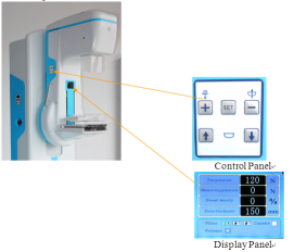
10. A total of 3 pieces of large size full color LCD screen display, operation table 8 inch LCD screen is a touch key.
11. Comfortable Compression:
When some degree of pressure is required for radiography, it allows you to presser the appropriate pressure(up to a maximum of 20kg) and is equipped with MICOM Control’s Soft-touch system which is designed to minimize the discomfort of the examine with in the pressure range.
Tissue Compression: Manual and Motorized (Max 20kg)
Compression Force and Thickness Data Display
Micro Control’s Compression
Automatic Release
12. Optional Intelligent Automatic Exposure Control(AEC)
With the Automatic Exposure Control system, it is possible to produce images with reliable intensity suitable for and film, screen, or method of radiography.
Furthermore, it greatly enhances the convenience of radiography by embedding the Full-AEC function which is capable of utilizing the Auto kV
Type: Solid-State Detector
Microprocessor Control
AEC Mode: Full AEC(Auto kV)
Semi AEC (kV Select)
Manual (kV, mAs Select)
Density Adjustment: 16 density steps
V.Diagram
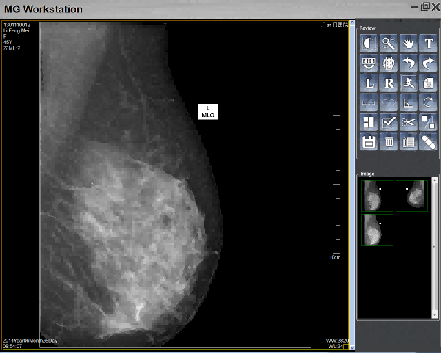
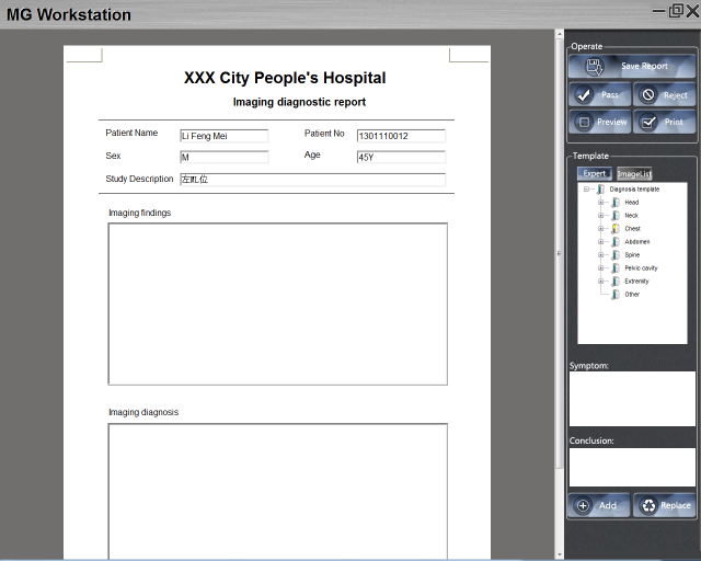
VI.Digital workstation:
|
|
Image Processing: Negative, Zoom, Roam, Select, Mirror, Counterclockwise rotation, Clockwise rotation, Typesetting image, Cut out, Report Map and so on |
Digital workstation
System introduction:
1.Login, Main interface
2.Image Processing
3.Reporting Interface
4.Statistics
5.System Management
*More details refer to User Manual